
PhasorPy#
PhasorPy is an open-source Python library for the analysis of luminescence lifetime and hyperspectral images using the phasor approach.
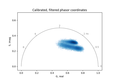
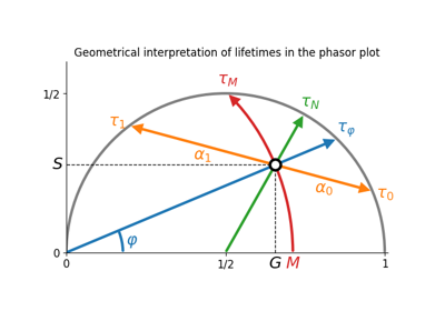
The phasor approach represents time-resolved and spectral signals as phasor coordinates (normalized Fourier coefficients at harmonic frequencies) for intuitive visualization and analysis.
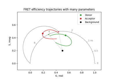
PhasorPy enables reproducible phasor-based fluorescence lifetime imaging (FLIM) and hyperspectral imaging (HSI) workflows in scientific Python. It provides tools to read microscopy data in many file formats and to calculate, calibrate, filter, visualize, and interconvert phasor coordinates, lifetimes, and signals. Phasor coordinates can be exported to standard formats and analyzed through cursor-based region-of-interest selection, cluster detection, multi-component unmixing, FRET efficiency and concentration estimation.
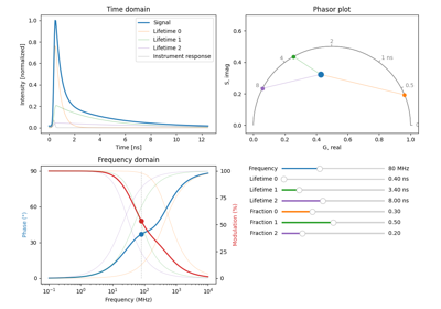
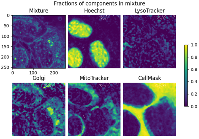
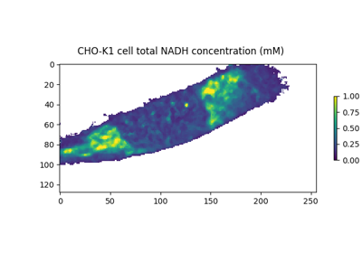


Documentation#
The PhasorPy documentation thoroughly documents all aspects of the library, including:
Other versions: latest development, all
Resources#
Source code is maintained on GitHub.com.
Releases are published on PyPI.org and conda-forge.
Data files are shared on Zenodo.org.
Ask questions and report issues on the GitHub issue tracker.
News#
July 20, 2026: PhasorPy 0.12 is released.
July 16, 2026: osx-arm64 packages are available for conda-forge.
July 2, 2026: 300th commit to the PhasorPy source code.
June 6, 2026: PhasorPy 0.11 is released.
May 12, 2026: PhasorPy 0.10 is released.
Jan 1, 2026: PhasorPy 0.9 is released.
Oct 10, 2025: PhasorPy 0.8 is released.
Aug 22, 2025: PhasorPy 0.7 is released.
Aug 16, 2025: The new PhasorPy logo is live.
June 22, 2025: PhasorPy 0.6 is released.
June 16, 2025: 200th commit to the PhasorPy source code.
Apr 11, 2025: PhasorPy 0.5 is released.
Jan 30, 2025: PhasorPy 0.4 is released.
Jan 25, 2025: The liffile library for reading Leica LIF files is released.
Jan 8, 2025: PhasorPy is available on conda-forge.
Dec 16, 2024: PhasorPy 0.3 is released.
Nov 30, 2024: PhasorPy 0.2 is released.
Oct 22, 2024: PhasorPy project update and practical are presented at the 18th LFD Workshop.
Sept 30, 2024: PhasorPy 0.1 is released.
Sept 23, 2024: 100th commit to the PhasorPy source code.
June 12, 2024: PhasorPy is featured at the CZI Open Science 2024 meeting.
Feb 16, 2024: First tutorial added to the documentation.
Nov 1, 2023: The ptufile library for reading PicoQuant PTU files is released.
Oct 26, 2023: The PhasorPy project is introduced at the 17th LFD Workshop.
Oct 5, 2023: The PhasorPy community on Zenodo is used to share data files.
Sept 1, 2023: Bruno Pannunzio joins the PhasorPy project.
Aug 22, 2023: The library documentation is published at www.phasorpy.org.
Aug 21, 2023: First PhasorPy developer meeting, to be held remotely every two weeks.
July 6, 2023: PhasorPy’s organization and repository on GitHub are live.
July 1, 2023: Christoph Gohlke joins the PhasorPy project.
Sept 2022: A grant from the Chan Zuckerberg Initiative is awarded to Leonel Malacrida and Enrico Gratton for the development of the PhasorPy library and community.

Events#
The PhasorPy project is presented at the following events:
Bruno Pannunzio. PhasorPy and napari-phasors: open source tools for FLIM-phasor analysis. F-Techniques Worskhop Series: FLIM. January 13-15, 2026. Portuguese Platform of BioImaging (PPBI), Portugal.
Bruno Schüty. PhasorPy hands-on training. LFD/AIM Workshop on Fluorescence Advanced Imaging Research (FLAIR). January 12-15, 2026. University of California, Irvine, USA.
Bruno Pannunzio and Leonel Malacrida. The PhasorPy library for FLIM and HSI phasor analysis. 6th Workshop in Advanced Microscopy and Biophotonics. November 23-28, 2025. Institut Pasteur de Montevideo, Uruguay.
Leonel Malacrida. PhasorPy library for open-source phasor analysis of FLIM and HSI: present, future and some applications in biological fluorescence. 12th International Weber Symposium. June 15-20, 2025. Genoa, Italy.
Bruno Pannunzio, Bruno Schuty Teske, Michelle Digman, Christoph Gohlke, and Leonel Malacrida. PhasorPy: an open-source Python library for the analysis of fluorescence lifetime and hyperspectral images using the phasor approach. 69th Annual Meeting of the Biophysical Society. February 15-19, 2025. Los Angeles, USA.
Leonel Malacrida. Introduction to PhasorPy. Third LFD around the world meeting. December 9, 2024. Online.
Leonel Malacrida. The PhasorPy library for FLIM and HSI phasor analysis. Optica Webinar Series. November 25, 2024. Online. Recording.
Bruno Schüty. Phasor Analysis in fluorescence lifetime imaging and spectral imaging with PhasorPy. GloBIAS Bioimage Analysts Workshop. November 5-8, 2024. Gothenburg, Sweden. Recording.
Bruno Pannunzio. PhasorPy: an open-source Python library for the analysis of fluorescence lifetime and hyperspectral images using the phasor approach. Virtual I2K 2024: Online tutorials on image analysis. October 29, 2024. Online. Recording.
Leonel Malacrida. Unlocking the phasor approach for FLIM and HSI: Insights from PhasorPy, a comprehensive Python library for phasor plot analysis. Euro-Bioimaging Virtual Pub. October 25, 2024. Online. Recording.
Bruno Schüty. PhasorPy hands-on training. 18th LFD Workshop in Advanced Fluorescence Imaging and Dynamics. October 21-24, 2024. Laboratory for Fluorescence Dynamics, University of California, Irvine, USA.
Christoph Gohlke. Introduction to PhasorPy. 18th LFD Workshop in Advanced Fluorescence Imaging and Dynamics. October 21-24, 2024. Laboratory for Fluorescence Dynamics, University of California, Irvine, USA.
Bruno Pannunzio, Bruno Schüty, Christoph Gohlke, and Leonel Malacrida. PhasorPy: un nuevo software abierto para el análisis de imágenes espectrales y tiempo de vida por gráficos de fasores. Semana Académica del Hospital de Clínicas. September 23-27, 2024. Montevideo, Uruguay.
Leonel Malacrida. Phasor analysis for FLIM & STED. Montreal Light Microscopy Course (MLMC). In-Focus: STimulated Emission Depletion (STED) Microscopy. July 22-26, 2024. Montreal, Canada.
Leonel Malacrida. The phasor approach for biophysical studies of condensates and membranes using DAN probes. XXVII Congreso Nazionale SIBPA. June 16-20, 2024. Genoa, Italy.
Leonel Malacrida. PhasorPy: a Python open-source library for phasor analysis of FLIM and HSI data. CZI Open Science 2024. June 11-14, 2024. Boston, USA.
Leonel Malacrida. The phasor plots for time-resolved and hyperspectral imaging: fundamentals and new developments. Sociedad Argentina de Microscopia. May 29-31, 2024. Tucuman, Argentina.
Leonel Malacrida. The phasor plot analysis for hyperspectral imaging and time-resolved imaging: PhasorPy developments and its applications. Latin American Hub for Bioimaging Through Open Hardware (LIBRE Hub) Seminars. May 15, 2024. Online. Recording.
Leonel Malacrida. PhasorPy: a Python open-source library as SimFCS legacy for phasor analysis of FLIM and HSI data. Frontiers in Biological Fluorescence 2024. May 10, 2024. University of California, Irvine, USA.
Leonel Malacrida. Microscopía no lineal en Uruguay: 2P-FLIM y microscopio DIVER para estudiar procesos in vivo en la profundidad del tejido. Simposio en Microscopía y Bioimágenes: avances y desafíos. April 4-5, 2024. University of the Republic (UdelaR). Montevideo, Uruguay.
Leonel Malacrida. Microscopy techniques applied to the analysis of biosensors performance. 1st Latin-American Workshop: Development and applications of biosensors: from fluorescent proteins to synthetic biology. Nov 27 - Dec 1, 2023. Institut Pasteur de Montevideo, Uruguay.
Bruno Schüty, Bruno Pannunzio, and Leonel Malacrida. Practical section 3: PhasorPy and Napari plugin for FLIM & HSI analysis. 5th Workshop in Advanced Microscopy and Biophotonics. November 19-24, 2023. Institut Pasteur de Montevideo, Uruguay.
Christoph Gohlke and Leonel Malacrida. Introduction to PhasorPy. 17th LFD Workshop in Advanced Fluorescence Imaging and Dynamics. October 23-27, 2023. Laboratory for Fluorescence Dynamics, University of California, Irvine, USA.
Publications#
Publications using PhasorPy:
González-Gutiérrez M, Vázquez-Enciso DM, Mateos N, Hwang W, Torres-García E, Hernández HO, Chacko JV, Coto Hernández I, et al. PSF-driven spatio-temporal blending in fluorescence lifetime imaging microscopy and its mitigation via mean-shift super-resolution-based masking. bioRxiv (2026)
Schuty B, Garcia MJ, Khuon S, Malacrida L. Phasor analysis of RGB camera data enables fluorescence microscopy unmixing and brightfield segmentation in a commercial microscope. Sens Bio-Sens Res. 52: 101014 (2026)
Prieto D, Rehermann MI, Fabbiani G, Vitar M, Trujillo-Cenoz O, Falco MV, Cuparo M, Trigo FF, et al. A filopodia-based dendritic mechanosensory compartment in CSF-contacting neurons. bioRxiv (2026)
Halbers LP, Brennan CK, Scipioni L, Tedeschi G, Torrey ZR, Parag-Sharma K, Labra B, Gohlke C, et al. Expanded applications of bioluminescence microscopy with phasor analysis. Cell Rep Methods. 6(4): 101344 (2026)
Fuller EB, Cole KH, Halbers LP, Chan CET, Ng KK, Scipioni L, Gohlke C, Digman MA, et al. Bioluminescent probes for multiplexed RNA imaging. J Am Chem Soc. 148(11): 11521-11530 (2026)
Sabri E, Mangiarotti A, Dimova R. Fluorescence-based mapping of condensate dielectric permittivity uncovers hydrophobicity-driven membrane interactions. Nat Commun. 17:3155 (2026)
Pannunzio B, Cespedes P, Diaz M, Ali D, Rial A, Malacrida L. High-throughput single-cell spectroscopy using phasor analysis of spectral flow cytometry. bioRxiv (2026)
Zhao W, Samimi K, Skala MC, Datta R. FLIM playground: an interactive, end-to-end graphical user interface for analyzing single cells with fluorescence lifetime imaging microscopy. bioRxiv (2025)
Sabri E, Mangiarotti A, Schmitt CNZ, Dimova R. Seeing the chemistry of biomolecular condensates: in situ mapping of composition and water content. bioRxiv (2025)
Publications mentioning PhasorPy:
Lace B, Krohn P, Batzenschlager M, Fieß V, Molina D, Ragni L, Ott T. Fluorescence lifetime imaging in plants: practical guidelines for multiplexing, label-free imaging and data analysis. bioRxiv (2026)
Harkhoe R, Ferrari G, Dmitriev RI, Kuhl M. Luminescence lifetime imaging: key concepts and advances in the image acquisition, processing, and analysis pipeline. ACS Meas Sci Au. acsmeasuresciau.5c00209 (2026)
Georgakoudi I, Skala MC, Quinn KP, Stringari C, Sorrells JE, Heikal AA, Li LZ, Xu HN, et al. Consensus guidelines for cellular label-free optical metabolic imaging: ensuring accuracy and reproducibility in metabolic profiling. J Biomed Opt. 30(S2): S23901 (2025)
Wetzker C, Zoccoler ML, Iarovenko S, Okafornta CW, Nobst A, Hartmann H, Muller-Reichert T, Haase R, et al. A fluorescence lifetime separation approach for FLIM live-cell imaging. J Microscopy. 301(1): 91-106 (2025)
Wetzker C, Zoccoler ML, Iarovenko S, Okafornta CW, Nobst A, Hartmann H, Müller-Reichert T, Haase R, et al. Fluorescence lifetime unmixing: a new workflow for FLIM live-cell imaging. bioRxiv (2025)
Michalet X. AlliGator: open source fluorescence lifetime imaging analysis in G. bioRxiv (2025)
Kapsiani S, Laubli NF, Ward EN, Shehata M, Kaminski CF, Kaminski Schierle GS. FLIMPA: a versatile software for fluorescence lifetime imaging microscopy phasor analysis. Anal Chem. 97(22): 11382-11387 (2025)
Vallmitjana A, Torrado B, Durkin AF, Dvornikov A, Rajil N, Ranjit S, Balu M. GSLab: open-source platform for advanced phasor analysis in fluorescence microscopy. Bioinformatics. 41(4): btaf162 (2025)
Weber F, Iskrak S, Ragaller F, Schlegel J, Plochberger B, Sezgin E, Andronico LA. VISION - an open-source software for automated multi-dimensional image analysis of cellular biophysics. J Cell Sci. 137(20): jcs262166 (2024)
Mangiarotti A, Dimova R. Chapter five - The spectral phasor approach to resolving membrane order with environmentally sensitive dyes. Methods Enzymol. 700:105-126 (2024)
Cite#
Please cite doi: 10.5281/zenodo.13862586 if PhasorPy contributes to research that leads to a publication.
Contact#
PhasorPy is a community-maintained project.
Contributions in the form of bug reports, bug fixes, feature implementations, documentation, datasets, and enhancement proposals are welcome.
Report issues and ask questions about PhasorPy on the GitHub issue tracker.
Alternatively, contact the PhasorPy developers directly.